Some diagnoses are made not by a single test but by recognising a pattern of cells, and the granuloma is a classic example.
What happened
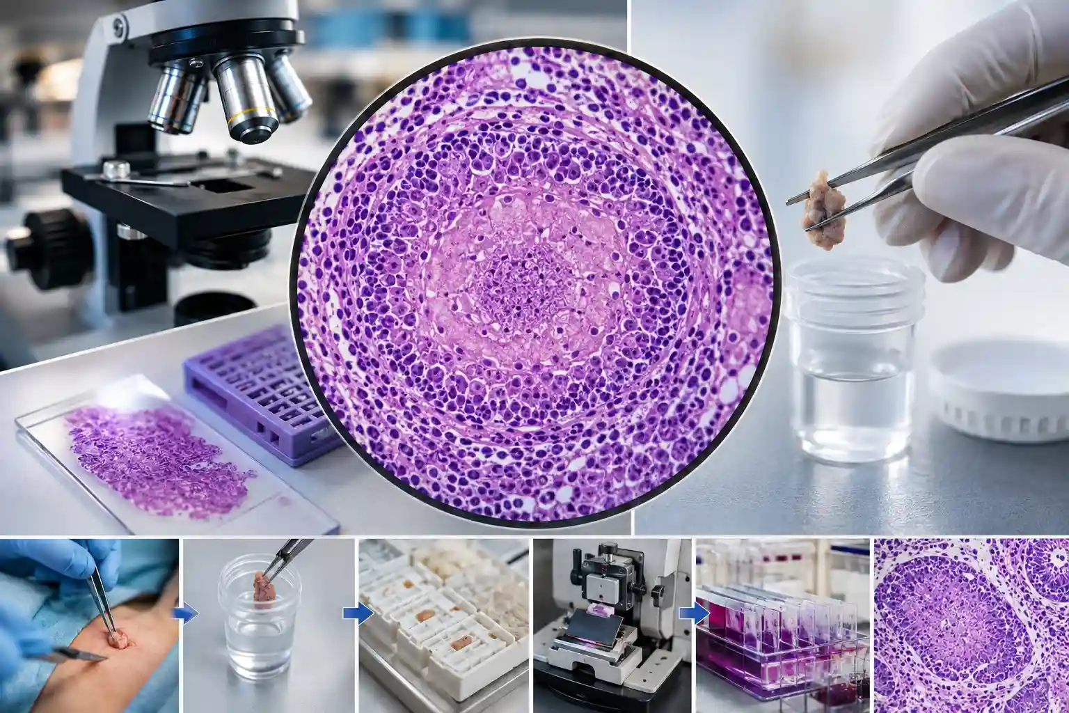
When the body meets something it cannot easily clear, certain infections, or conditions such as sarcoidosis, immune cells can organise into a compact, rounded nodule called a granuloma. Under the microscope, a pathologist recognises its ring of “epithelioid” immune cells and, sometimes, large multinucleate giant cells. In tuberculosis, the centre may show “caseating” (cheese-like) necrosis, a strong clue to the cause.
Why it mattered
Spotting a granuloma on a biopsy narrows a wide list of possibilities and guides the next step, for example, molecular testing to confirm tuberculosis, or correlation with the clinical picture for other causes. It is a good example of how histopathology and molecular testing complement each other: the microscope raises the question, and the right follow-up test helps answer it.
Key points
- A granuloma is an organised collection of immune cells the body forms to “wall off” a trigger
- Caseating (necrotic-centred) granulomas are characteristic of tuberculosis
- Other causes include sarcoidosis, some fungal infections, and reactions to foreign material
- The finding guides confirmatory testing and clinical correlation, rather than standing alone
How this connects to our testing
Recognising patterns like this is the everyday work of histopathology, PathMole examines biopsies from small samples to major resections, and can point toward the right follow-up, such as molecular Tuberculosis (TB) testing. See the Test List, or contact the lab to discuss sample requirements. Pricing is shared on enquiry.
Sources
Standard histopathology references on granulomatous inflammation (epithelioid and giant-cell reaction; caseating necrosis in tuberculosis). · World Health Organization: tuberculosis. See also our explainer on the WHO Classification of Tumours.